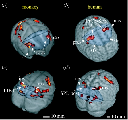
Figure 7.
Comparison of saccade-related activity in monkeys and humans. (a,c) Monkey and (b,d) human brain images are cut to show activity buried in the sulci in the frontal (a,b) and posterior parietal (c,d) cortices. Asterisks and crosses indicate the regions showing the highest selectivity to the contraversive saccades in the frontal and parietal cortices, respectively. FEF, frontal eye field; LIPd, dorsal lateral intraparietal; PrCS/SFS, intersection of the precentral sulcus and the superior frontal sulcus; SPL, superior parietal lobule; as, arcuate sulcus; ips, intraparietal sulcus; prcs, precentral sulcus; sfs, superior frontal sulcus; post, posterior. Adapted from Koyama et al. (2004).

