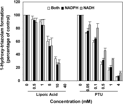
Fig. 5.
Inhibition of triazolam 1-hydroxylation in HLMs by α-lipoic acid and PTU. Triazolam 1-hydroxylation activities in HLMs were measured in the absence and presence of increasing concentrations of α-lipoic acid (left) or PTU (right). Cofactors included either NADH alone (black bar), β-NADPH alone (hatched bar), or a mixture of NADH plus β-NADPH (open bars). Results are expressed as the percentage of control activities and represent the mean ± S.D. of four experiments using HLMs from four different donors. *, P < 0.05 by Student-Newman-Keuls test for NADH alone incubation versus β-NADPH alone and NADH/β-NADPH incubations measured at the same inhibitor concentration. Similar results were obtained for 4-hydroxylation of triazolam (data not shown).

