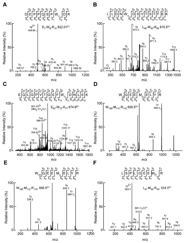
Figure 1. Identification of modified apoAI in human atheroma by mass spectrometry.

CID spectra were acquired after direct or in gel tryptic digest of imunnoaffinity purified apoAI derived from human atheroma. Doubly charged ions were detected and fragmented in an LC-tandem mass spectrometry experiment. A. Peptide D1-R10 containing monohydroxytryptophan at residue 8. B. Peptide L46-K59 containing monohydroxytryptophan at residue 50. C. Peptide E62-K77 containing monohydroxytryptophan at residue 72. D. Peptide W108-R116 containing monohydroxytryptophan at residue 108 and methionine sulfoxide at residue 112. E. The same peptide as in D, but the tryptophan at residue 108 is converted to dihydroxytryptophan. F. Peptide L41-R49 containing methionine sulfoxide as at residue 48.
