
Figure 2. Aminoadipic acid levels in plasma and lesion apoAI.
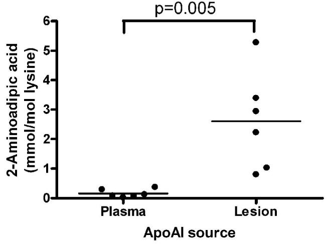
ApoAI was isolated by immunoaffinity chromatography from the plasma of six healthy subjects and from six atheroma samples. 2-aminoadipic acid levels were quantified by mass spectrometry after acid hydrolysis, and normalized to apoAI lysine content. Data show mean of duplicate determinations for each sample (p=0.005 by two tailed t-test).