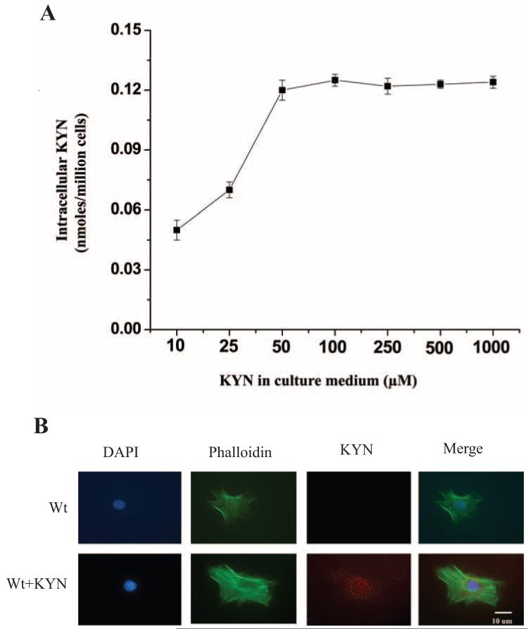
Figure 5.
Effect of exogenous KYN on intracellular KYN and KYN modification in mLECs. (A) mLECs from Wt were incubated with 0 to 1.0 mM KYN for 3 days. Cells were lysed, protein precipitated, and KYN estimated in the protein-free supernatants by HPLC. Results are the mean ± SD of results in three independent experiments. (B) Wt mLECs were incubated with 50 μM KYN for 3 days and immunostained for KYN-modified proteins. KYN-modified proteins (red) were detected in KYN-treated mLECs but not in untreated cells. Cells were also stained for F-actin by phalloidin (green) and nuclei with DAPI (blue). Images are representative of three independent experiments.

