Fig. 5.
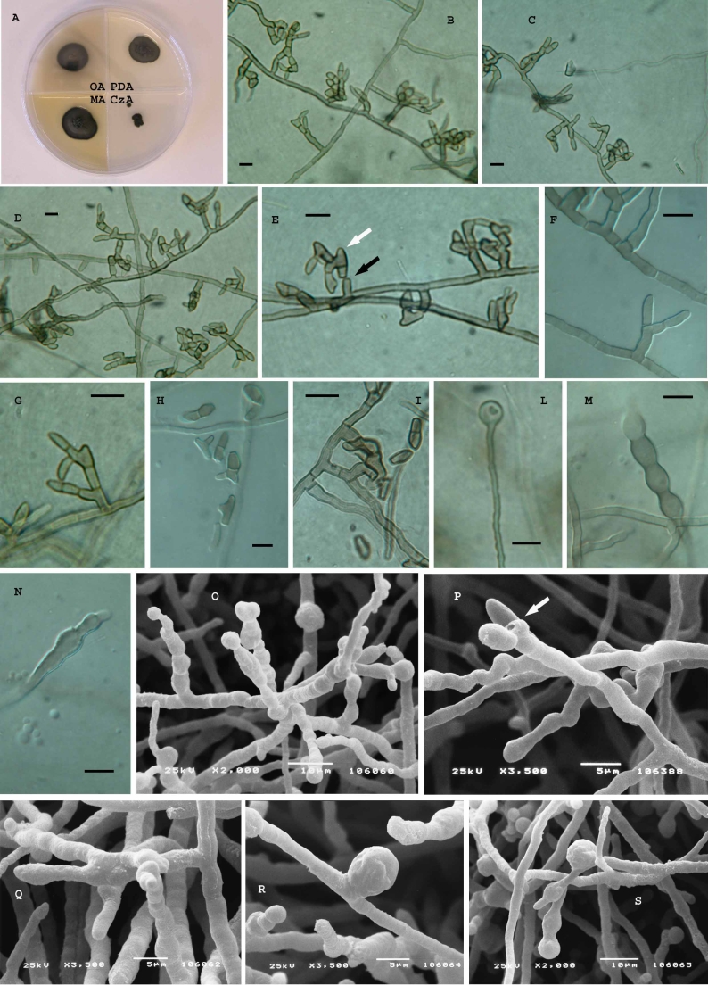
Recurvomyces mirabilis, CBS 119434 (= CCFEE 5264). A. Strain grown on different media after two mo of incubation at 15 °C. B–D. Hyphae with branched and unbranched conidiophores producing 0–1 septated conidia. E. Curved branched conidiophores schyzolytically seceding (black arrow) showing enteroblastic elongation (white arrow) at the apex. F, G. high magnification of branched conidiophores producing 1-celled conidia. H, I. 1- and 2-celled conidia and ramoconidia. L. Terminal swelling cell. M, N. swelling hyphae showing enteroblastic elongation. O, P. Unbranched conidiophores producing 1-celled conidia, scar is visible after schizolytic secession (P, arrow). Q. Branched conidiophore. R, S. swelling cell at intercalary (R) and terminal position (S). B–N. Light microscopy; Scale bars = 10 μm. O–S. SEM.

