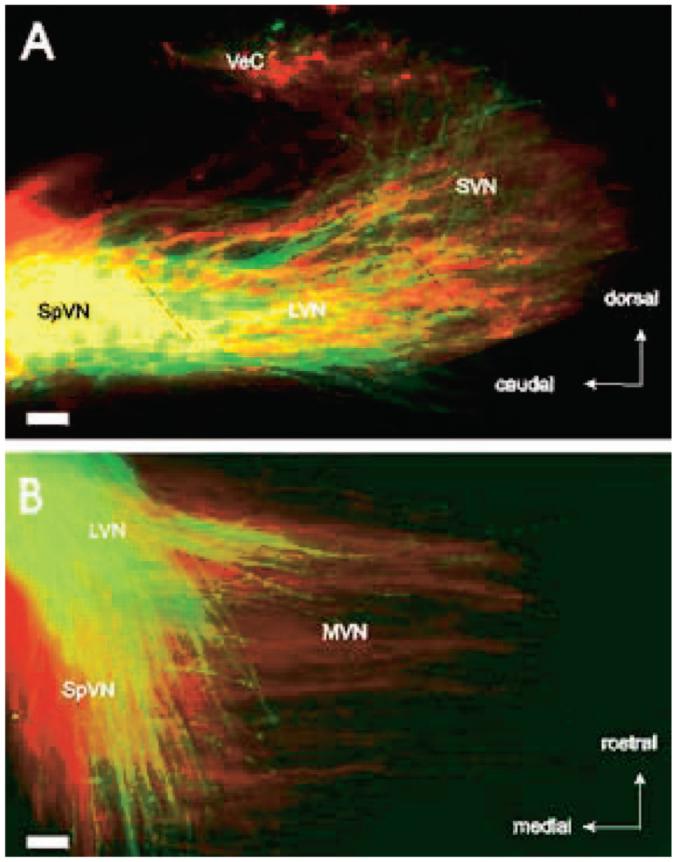
Figure 3.

Superimposed branching patterns of posterior vertical canal (A) and saccular (B) axons in the vestibular nuclei of fetuses in flight (green) and control (red) conditions. (A) Parasagittal sections show afferent axons from the posterior vertical canal project to overlapping vestibular areas in both control and microgravity-exposed fetuses. (B) Horizontal sections demonstrate that afferent axons from the sacculus project more medially in control than microgravity-exposed fetuses. LVN = lateral vestibular nucleus; MVN = medial vestibular nucleus; SpVN = spinovestibular nucleus; SVN = superior vestibular nucleus; VeC = vestibulocerebellum. Bar = 100 μm.
