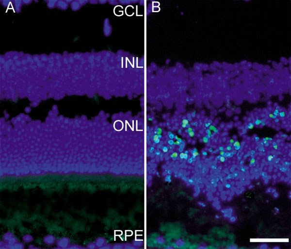
Figure 1.

Fluorescence photomicrographs of TUNEL-labeled mouse retinas. Control mice did not have bright light exposure (A). Experimental mice were sacrificed 28 h after light exposure (B). All nuclei are labeled with DAPI (blue). TUNEL-positive photoreceptors appear blue-green in B. Retinal layers are abbreviated as follows: retinal pigment epithelium (RPE), outer nuclear layer containing photoreceptor nuclei (ONL), inner nuclear layer (INL), and ganglion cell layer (GCL). Scale bar indicates 50 μm.
