
Fig. 4.
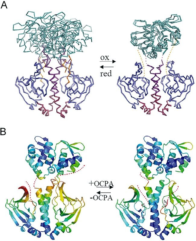
Dynamic information obtained from CprK crystal structures.
A. Overlay of the different CprK ligand-free structures. The left panel displays an overlay of the D. hafniense CprKC200S and D. dehalogenans CprK (PDB code 2H6C) depicted in ribbons coloured coded according for Fig. 1. The right panel displays a similar overlay but for the oxidized D. hafniense CprK structure.
B. Comparison between the CprKC200S ligand-free (to the left) and OCPA-soaked CprKC200S (to the right) structures. The ribbon models are coloured according to Cα B-factors.