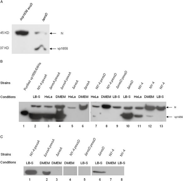
Fig. 7.

A. Western blot showing the specificity for the polyclonal antibody against Vp1656 for the ΔexsD strain (right lane) and the Δvp1656 exsD strain (left lane). A non-specific protein band (‘N’) was detected with this antisera and serves as a positive detection control in these experiments.
B. Expression of Vp1656 after bacteria were grown for 2 h in different conditions. Lane 1 shows purified protein Vp1656-6xHis.
C. Secretion of Vp1656 after bacteria were grown for 2 h in different conditions.
