
Fig. 5.
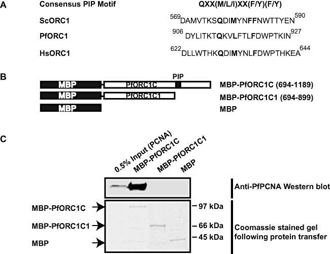
Identification of conserved PIP motif in ORC1.
A. The consensus PIP motif is shown on the top. The putative PIP domains present in ScORC1, PfORC1 and HsORC1 are also shown. The conserved residues are marked in bold.
B. The schematic diagrams of C-terminal region of PfORC1 with (PfORC1C) or without (PfORC1C1) PIP domain fused to MBP. The residues of PfORC1 taken in each construct are also shown. The PIP motif is marked as black box.
C. Pull-down experiments using MBP-fused ORC1 constructs. Two microgram of His6-PCNA was incubated with equal amount of MBP beads containing MBP-ORC1C (+PIP motif) or MBP-ORC1C1 (−PIP motif) or MBP alone followed by stringent washing of the beads using buffer as mentioned in the Experimental procedures. The bound proteins were released by boiling in SDS-PAGE loading buffer followed by Western blot analysis using anti-PfPCNA antibodies. The input lane shows 0.5% PCNA. The bottom panel shows the loading control for pull-down experiments with MBP alone or other fusion proteins.