Figure 1.
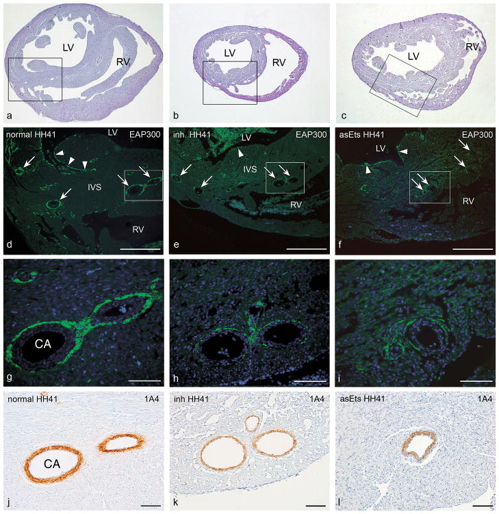
Photomicrographs of representative transverse sections of a normal control quail heart (HH41; a, d, g, and j ), a quail heart with a mechanical inhibition of epicardial outgrowth (inh; HH41; b, e, h, and k, and a chicken heart with hampered EPDC differentiation due to retrovirally induced down-regulation of Ets-1 and Ets-2 (asEts; HH41; c, f, i, and l). Panels a–c show haematoxylin-eosin stained overviews of the sections, at approximately mid-ventricular level. Boxed areas in a–c delineate the regions depicted in d–f. Serial sections were stained for Purkinje fiber cells with the EAP-300 antibody (in green) and nuclei were stained with DAPI (in blue). In the control heart, (d, g) normal development of the periarterial (arrows) and subendocardial (arrowheads) Purkinje fiber cells was observed. In contrast, in the inhibition embryo (e, h), and in the asEts-1/2 embryo (f, i), we observed a decrease in Purkinje fiber cell numbers. The details (g–i; boxed areas in d–f) show periarterially located Purkinje fiber cells. In embryos with disturbed epicardial contribution EAP-300 positive cell numbers were dramatically reduced compared to normal embryos. Quantification of the area occupied by periarterial Purkinje fibers was performed as described in the text under Materials & Methods, in sections like these, comparing coronary arteries of similar diameter. Panels j–l show the 1A4 (smooth muscle actin) staining in consecutive sections of g– i. The smooth muscle cell layer of the coronary arteries (CA) was slightly thinner in this mechanically PEO-inhibited embryo (k). However, this was not a consistent finding. Also the asEts-1/2 embryos displayed coronary arteries with normal thickness of the smooth muscle cell layer (l). CA, coronary artery; IVS, interventricular septum; LV, left ventricle; RV, right ventricle. Scale bars, 250 μm in d–f; 50 μm in g–l.

