Figure 3.
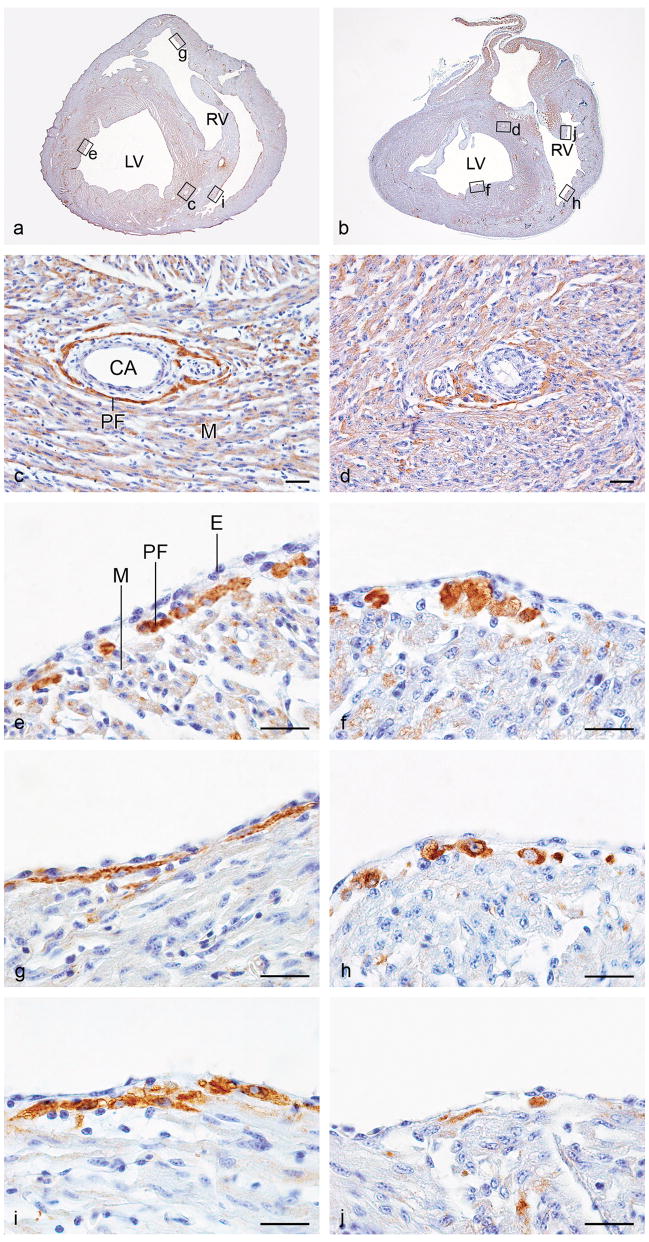
Photomicrographs of representative transverse sections of a normal control quail heart (HH 41; a, c, e, g, and i) and a quail heart with mechanical inhibition of epicardial outgrowth. (inh; HH 41; b, d, f, h and j). Sections were stained with immunofluorescent EAP-300 antibody and DAB-converted as described in the Materials & Methods section (c–j). Panels a and b show the complete sections at approximately mid-ventricular level, where coronary arteries of similar diameter are present. The location of the enlargements in panels c–j is indicated. In the mechanically inhibited heart, the periarterial EAP-300 signal is faint and diffuse (d) when compared to the signal in the periarterial Purkinje fibers in the normal control heart (c). This is consistent with the finding that EAP-300 immunofluorescence did not surpass background in these embryos (Figure 2). Additionally, EAP-300 staining revealed abnormally large rounded cells in the subendocardial compartment of the peripheral conduction system. These cells were not as neatly organized in fibers as the EAP-300 positive cells in controls (e, f; left ventricular and g, h; right ventricular ventral free walls). Hyperplasia and absence of fiber-like organization of the subendocardial Purkinje fibers was observed throughout, and is illustrated here by EAP-300 staining in the moderator band of the right ventricle (i, j; lateral side of the moderator band). CA, coronary artery, E, endothelium; LV, left ventricle; M, myocardium; PF, Purkinje fiber; RV, right ventricle. Scale bars, 20 μm.

