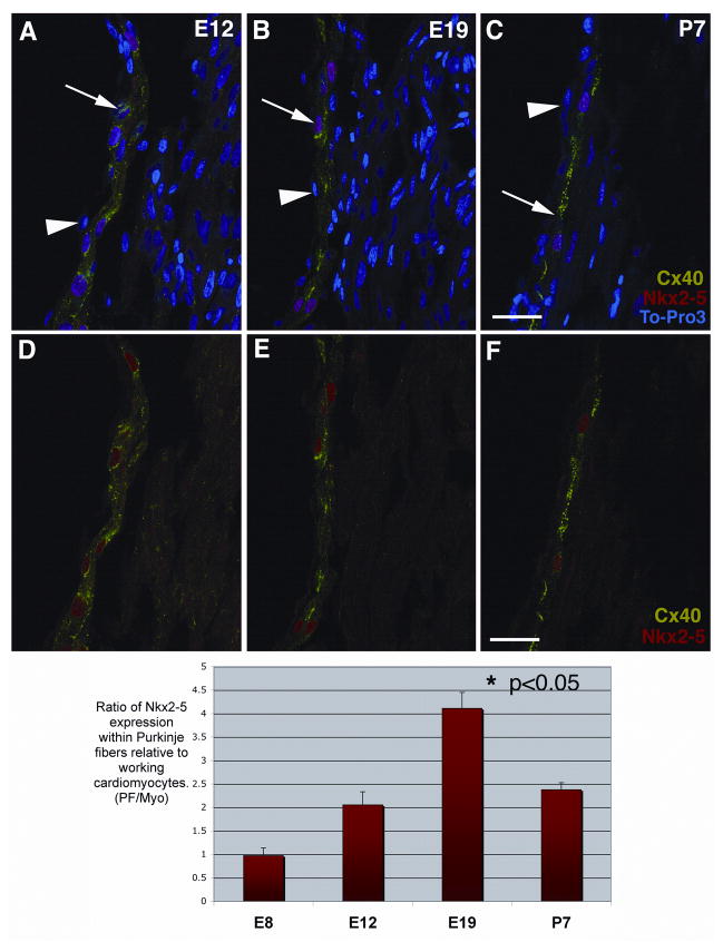
Figure 2. Increasing levels of nuclear-localized Nkx2-5 defines Purkinje fiber development.
Chick heart sections at four stages; E8, E12 (A, D), E19 (B, E) and P7 (C, F) were immunolabeled for Cx40 (shown in green, FITC), Nkx2-5 protein (shown in red, TRITC) and the nuclear dye, To-Pro3 (shown in blue). These laser scanning confocal images of chick heart sub-endocardium show that Nkx2-5 localizes mainly to the nuclei of Cx40-positive Purkinje fibers and to a lesser extent the nuclei of Cx40-negative cells in the working myocardium at E12, E19 and P7 (A, B, C). For clarity these same images are reproduced, excluding the nuclear stain (D, E, F). These single optical images were used to quantify Nkx2-5 immuno-positive nuclear signal within Purkinje fibers (Cx40-positive) and working cardiomyocytes (Cx40-negative) at the same three stages; E12, E19 and P7. We measured the mean nuclear intensity of the Nkx2-5 immuno-positive signal and expressed it as a ratio, Purkinje fiber/working cardiomyocyte. We found that at E19 the Nkx2-5 immuno-intensity ratio was 100% more than at E12 and also P7 (P< 0.05). The quantitative analysis of E8 cells was undertaken on sub-endocardial cardiomyocytes prior to Cx40 up regulation. This analysis was carried out on multiple images (n=20) from multiple animals (n=3). Scale bars 50μm.

