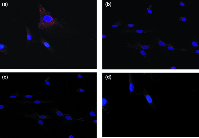
Fig. 4.
Analysis of fibronectin protein in myometrium and leiomyoma cells as analysed by cytoimmunofluorescence. Pre-treatment leiomyoma cells (a) demonstrated a higher amount of protein (higher red fluorescence) compared to myometrial cells (c). Decreased red fluorescence in post ATRA (10−7m) treated leiomyoma cells (b) indicated a decreased amount of fibronectin protein. Minimal decreased red fluorescence was observed in myometrial cells (d). Red fluorescence is indicative of fibronectin protein, DAPI (blue fluorescence) strongly binds to DNA and indicates the nucleus in the cell. Magnification: 40×.

