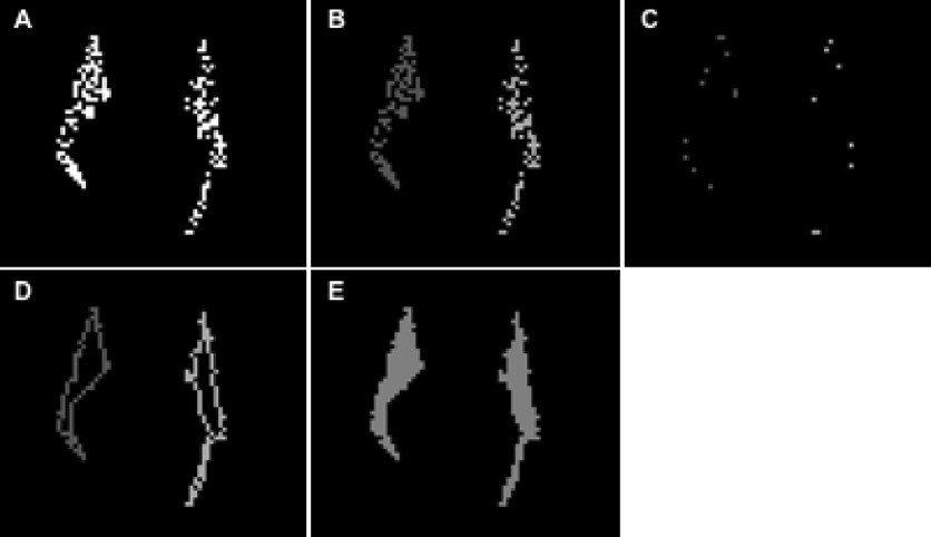
Figure 1.
A) Cross-section of the fibers of the forceps-minor with a coronal plane. The white dots represent voxels that belong to fiber-lines. These voxels are referred to in the text as “fiber-points”. B) The “fiber-points” of the forceps-minor that belong to the left and right hemispheres are assigned to different clusters. The cluster corresponding to the right hemisphere is shaded darker grey and the one corresponding to the left hemisphere is shaded lighter grey. C) The vertices of the minimum perimeter polygons that enclose all the “fiber-points” in each cluster. D) The complex lines that connect the “fiber-points” closer to the sides of the minimum perimeter polygons. E) The voxels that were eventually considered to be part of the volumetric representation of this fiber-bundle.

