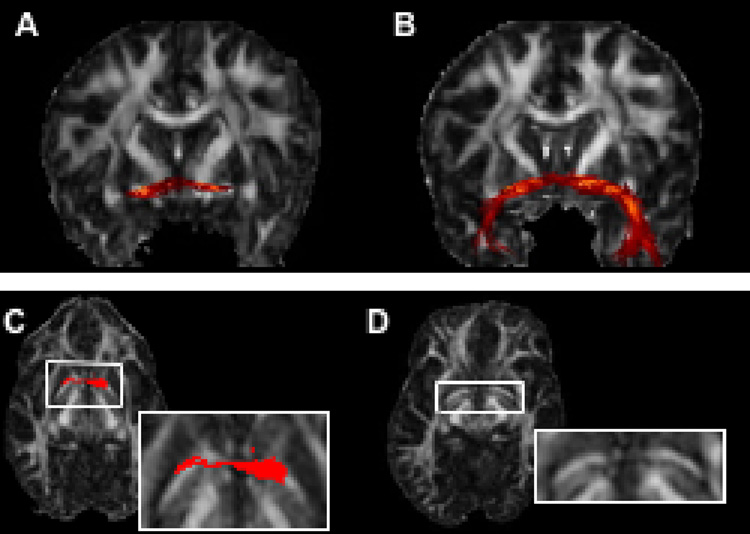
Figure 3.
Fibers of the AC, reconstructed from SE-EPI-DTI12 (before distortion correction) (A) and Turboprop-DTI (B) data from the same subject. Tracking based on SE-EPI-DTI12 data mapped only part of the AC (A). Tracking based on Turboprop-DTI data produced a more complete representation of the AC (B). The part of the AC that is included in the axial slice shown in C and D is characterized by increased curvature in SE-EPI-DTI12 (C), compared to Turboprop-DTI (D), due to magnetic susceptibility-related distortions. The cross-section of the traced AC fibers with this axial plane is shown in red. Although the curvature of the AC is increased due to the distortions, the diffusion orientation information within the AC remains the same. Thus, the estimated pathway was not curved enough during the fiber-tracking procedure, reached the walls of the bundle, and was terminated prematurely.

