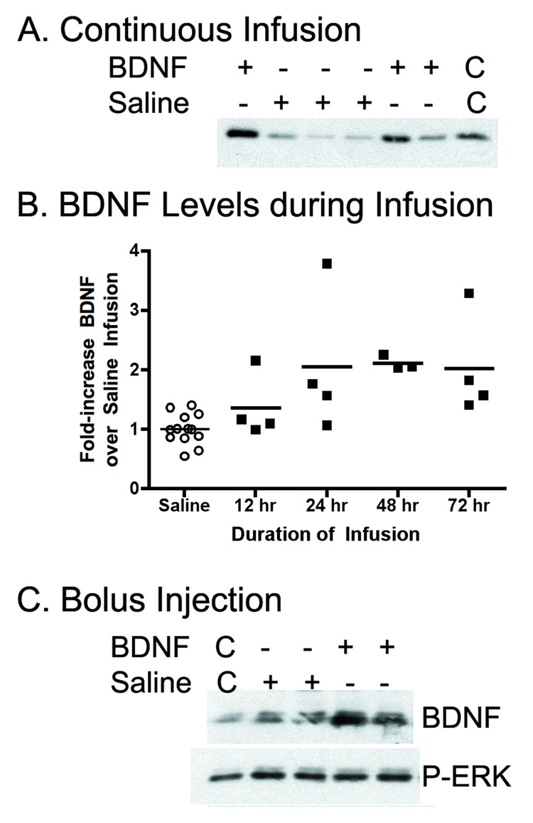
FIGURE 2.
A. Infusion of BDNF (2.5 µg per side over 72 hours) into the brain increased tissue levels of BDNF in the hippocampus. Linearity control lanes (C) contain double-volumes of one of the samples. B. Levels of BDNF in the hippocampi of individual rats during infusions of saline or BDNF reveal two-fold increases during BDNF infusions that was different from controls at 24, 48 and 72 hours (p<0.05). C. After bolus injections of BDNF, hippocampal BDNF levels increase, but active ERK (P-ERK) do not increase. Linearity controls (C) contain half-volumes of one of the samples.

