Fig. 1.
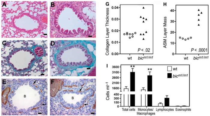
Mice deficient for bic/miR-155 show increased lung airway remodeling (A to F) Histological examination of sections of lung bronchioles from control wild-type (A, C, and E) and bicm1/m1 mice (B, D, and F). Scale bar, 100 μm. (A and B) Haematoxylin and eosin stain; (C and D) Masson Trichrome stain; (E and F) Immunohistochemical staining for smooth muscle actin. Collagen layer (white arrows), lung myofibroblasts (black arrows), bronchioles (B), and blood vessels (V) are indicated. (G) Quantitation of peribronchiolar collagen thickness or (H) airways smooth muscle cell (ASM) mass in bicm1/m1 mice compared with that of wild-type mice. (G) P < 0.02 or (H) P < 0.0001, in comparison with wild-type group, Student's two-tailed t test. Open circles, control mice; filled triangles, bicm1/m1 mice. Notably, bicm1/m1 mice with increased collagen layer thickness also had increased ASM mass. (I) Total and differential cell counts in BAL from the indicated mice. Data are the mean + SE from seven bic-deficient mice and six control mice. **P < 0.01 in comparison with wild-type group, Student's two-tailed t test.

