
Fig. 2.
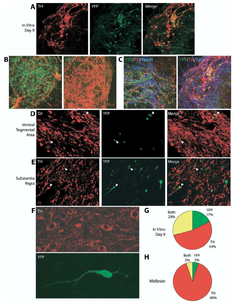
YFP reporter expression in differentiated THYFP/+ knock-in mES cells and adult THYFP/+ knock-in mouse midbrain. (A, B) THYFP/+ mES cells were differentiated in vitro for 9 days on PA6 cells into dopaminergic neurons and co-stained with antibodies to TH (red) and YFP (green). (C) THYFP/+ mES cells were differentiated for 9 days and co-stained for TH (red), YFP (green) and the nuclear neuronal marker NeuN (blue) or for TH (red), YFP (green) and the neuronal marker TuJ1 (blue). Triple overlap in expression appears purple. Cryosections of THYFP/+ heterozygous adult brain were co-stained with antibodies to TH (red) and YFP (green) in the ventral tegmental area (D) and substantia nigra (E). Examples of cells with overlapping expression are indicated by arrowheads. A higher magnification confocal stack image of neurons in the substantia nigra is seen in (F). (G) Quantification of differentiated TH and YFP expressing cells in vitro. Of the total cells (n=1508), 54% expressed TH alone (n=804), 17% expressed YFP alone (n=262), and 29% expressed both TH and YFP (n=442). (H) Quantification of TH and YFP expressing cells in the mouse midbrain. Of the total cells (n=426), 90% expressed TH alone (n=382), 5% expressed YFP alone (n=21), and 5% expressed both TH and YFP (n=23).