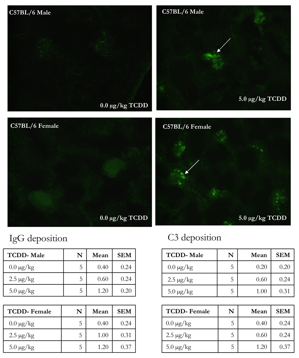
Figure 3.
The kidneys from 24 week-old C57BL/6 mice that were prenatally exposed to 0.0, 2.5 and 5.0 µg/kg TCDD were collected, fixed, section and stained with FITC-labeled anti-IgG. The above figures are representative of kidneys stained with FITC-anti-IgG from control female (A) and male (C) or 5.0 µg/kg TCDD-exposed female (B) and male (D) mice. The table data show the mean± SEM of the IgG and C3 disposition scores of 5 mice/treatment/gender.

