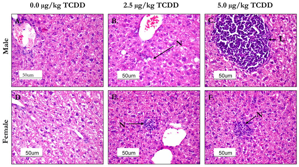
Figure 5.
The livers from 24 week-old C57BL/6 mice that were prenatally exposed to 0.0, 2.5 and 5.0 µg/kg TCDD were collected, fixed, section and stained with H&E stain. The above images are representative of control male (A) and female (D), 2.5 µg/kg TCDD male (B) and female (E) and 5.0 µk/kg TCDD male (C) and female (F) liver sections. Livers from male mice prenatally-exposed to 5.0 µg/kg TCDD showed infiltration of lymphocytes (L) round central veins compared to control group. Livers from the 2.5 µg/kg prenatally-exposed TCDD males and 5.0 µg/kg TCDD females contained foci of necrosis (N) with inflammatory cells not evident in the controls.

