
Fig.2.
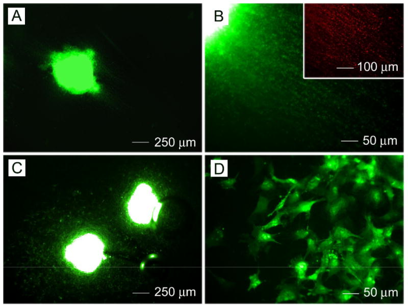
Fluorescence microscopy images of CE3 embryoid bodies after seeding onto (A, B) aligned and (C, D) randomly oriented PCL nanofibers for 14 days. Inset: staining with neuron marker Tuj1.
Official websites use .gov
A
.gov website belongs to an official
government organization in the United States.
Secure .gov websites use HTTPS
A lock (
) or https:// means you've safely
connected to the .gov website. Share sensitive
information only on official, secure websites.
Fluorescence microscopy images of CE3 embryoid bodies after seeding onto (A, B) aligned and (C, D) randomly oriented PCL nanofibers for 14 days. Inset: staining with neuron marker Tuj1.