
Figure 2.
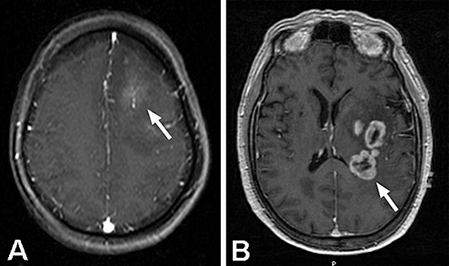
A. Post‐contrast, axial MRI of a patient with anaplastic astrocytoma (AA) that showed microscopic evidence of intravascular thrombosis in the biopsy specimen. The MRI shows mild contrast‐enhancement of the neoplasm (arrow) with surrounding T1‐hypointensity, typical of AA. B. Post‐contrast, axial MRI of a patient with glioblastoma, demonstrating the pattern of “ring‐enhancement” surrounding central necrosis that is characteristic of this disease (arrow).