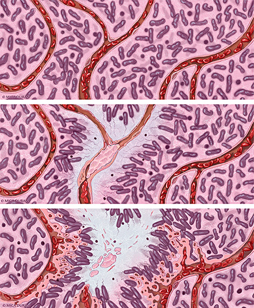
Figure 3.

Schematic representation of a proposed model of progression of anaplastic astrocytoma (AA; WHO grade III) to glioblastoma (upper panel). In AA, tumor cells infiltrate through central nervous system parenchyma, receiving oxygen and nutrients from intact native blood vessels (middle panel). A vascular insult occurs, causing endothelial injury, vascular leakiness and intravascular thrombosis. Thrombotic vaso‐occlusion leads to tumor cell migration away from hypoxia and toward a viable vasculature, creating a peripherally moving wave that is seen microscopically as pseudopalisading cells (lower panel). The zone of hypoxia and central necrosis expands, while hypoxic tumor cells of pseudopalisades secrete pro‐angiogenic factors (VEGF, IL‐8) that induce microvascular proliferation in adjacent regions, supporting an accelerated outward expansion of tumor. Illustration by Mica Duran.
