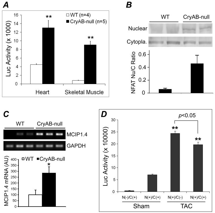
Figure 7.
CryAB suppresses NFAT transactivation in mice. A, NFAT-Luc transgenic mice were cross-bred with the KO (CryAB-null) mice and the luciferase (Luc) activities in myocardium and the soleus muscle of CryAB-null and wild type littermates (WT) were measured. Compared with WT, **: p < 0.01. B, Western blot analyses of NFATc4 in the nuclear and the cytoplasmic (Cytopla.) fractions of myocardium from WT and KO mice. A representative set of images are shown at the top and the nuclear to cytoplasmic NFAT ratios derived from the densitometry of the Western blot images are summarized in the bar graph. C, RT-PCR analyses of myocardial MCIP1.4 expression in KO mice. *: p < 0.05, KO vs WT. D, The NFAT-Luc mice were cross-bred with the CryAB TG mice and the resulting littermate mice with the indicated genotypes were subjected to TAC or sham surgery at 12 weeks. LV myocardial luciferase activities were assessed at 2 weeks after the surgery. N(-): NFAT-Luc Ntg; N(+): NFAT-Luc Tg; C(-): CryAB Ntg; C(+): CryAB Tg. Mean+SD; n = 4 mice/group; compared with either sham groups, **: p < 0.01.

