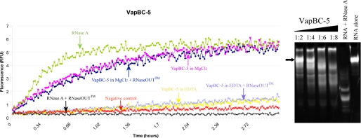
FIGURE 4.
In vitro ribonuclease activity of M. tuberculosis VapBC-5. A, fluorescence measurements as a function of time. A fluorescent substrate is incubated with VapBC-5 in different conditions. Fluorescence is measured when the substrate is cleaved which indicates the presence of ribonuclease activity. It clearly shows that VapC-5 activity is dependent on the presence of magnesium as shown by the magenta and navy blue curves. B, nuclease assay: polyacrylamide/urea denaturing gel showing from left to right:2 μm RNA stained by Sybr Green II with increasing concentration of VapBC-5 in MgCl2. The RNA was incubated for 5 h at 37 °C with 4, 8, 12, 16 μm VapBC-5, respectively, and the subsequent lanes shows the RNA incubated with RNase A as a positive control and the RNA alone. The black arrow points to the intact RNA. Degradation products appear (smears) when the RNA is incubated with VapBC-5.

