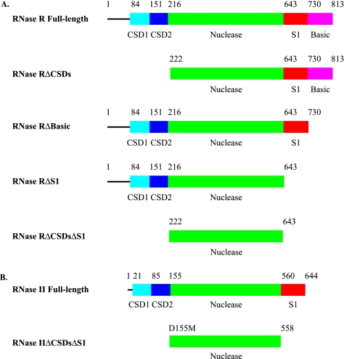
FIGURE 1.
Linear domain organization of RNase R and RNase II proteins. The CSDs are colored in cyan and blue for CSD1 and CSD2, respectively, the nuclease domains are in green, the S1 domains are red, and the low complexity, highly basic region, found in RNase R only, is in magenta. A, RNase R. RNase R full-length is the full-length wild-type RNase R protein. RNase RΔCSDs lacks both CSD1 and CSD2. RNase RΔBasic is missing the low complexity, highly basic region. RNase RΔS1 is missing both the S1 domain and the low complexity, highly basic region. RNase RΔCSDsΔS1 consists of the nuclease domain alone. B, RNase II. RNase II full-length is the full-length wild-type RNase II protein. RNase IIΔCSDsΔS1 contains the nuclease domain alone.

