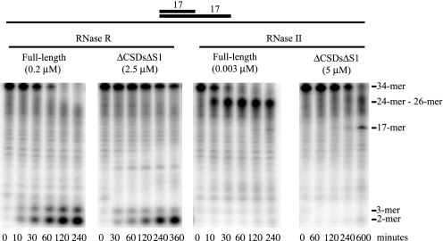
FIGURE 3.
Comparison of RNase II and RNase R full-length proteins and nuclease domain-truncated proteins on a substrate containing a duplex. Assays were carried out as described under “Experimental Procedures” with 10 μm ds17-A17 substrate and the indicated enzyme concentrations. Aliquots were taken at the indicated times and analyzed by denaturing PAGE. The origin of the band at the approximate position for an 8-mer that first appears at the 30-min time point with RNase RΔCSDsΔS1 is unknown. However, it is not observed reproducibly.

