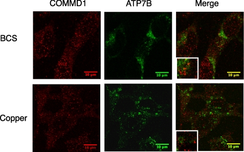
FIGURE 3.
Intracellular localization of COMMD1 and ATP7B in HepG2 cells following BCS or CuCl2 treatment. Confocal immunofluorescence microscopy of COMMD1 and ATP7B was performed after HepG2 cells are treated for 8 h with either 200 μm BCS or 50 μm CuCl2. Non-polarized HepG2 cells are shown; the pattern of COMMD1 in polarized cells was very similar.

