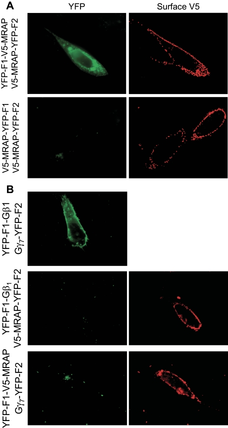
FIGURE 2.
Specificity of MRAP bimolecular fluorescence complementation. A, live CHO cells transfected with MRAP fused to YFP fragments on opposite sides or the same side of the MRAP protein. B, live CHO cells transfected with V5-MRAP-YFP-F2 and Gβ1-YFP-F1, YFP-F1-V5-MRAP, and Gγ7-YFP-F2, or Gβ1-YFP-F1 and Gγ7-YFP-F2 as shown. Left panels, YFP fluorescence. Right panels, surface expression of MRAP detected by staining live cells with anti-V5 antibody.

