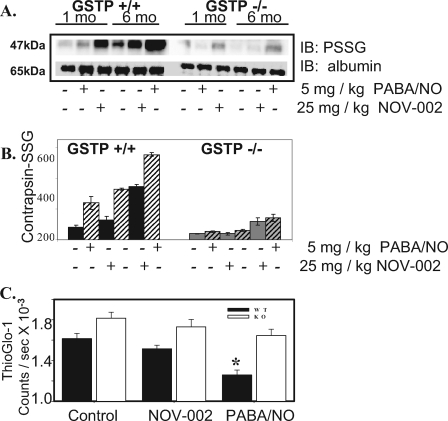
FIGURE 4.
S-Glutathionylation of serum and liver proteins in GSTπ WT and KO mice. Male GSTπ wild-type (GSTP1P2+/+) and knock-out (GSTP1P2-/-) mice (ages 1 and 3 months) were treated with an intravenous bolus of the oxidized glutathione mimetic, NOV-002 at 25 mg/kg or PABA/NO at 5 mg/kg. Blood was collected at various time points via orbital bleed. The plasma proteins (A) were separated by non-reducing SDS-PAGE and S-glutathionylated contrapsin was evaluated by immunoblot (IB) with PSSG antibody. The ratios of S-glutathionylated contrapsin to albumin were plotted in arbitrary units (B), solid, 1-month-old; hatched, 3-month-old animals (6 animals per treatment group). Free protein sulfhydryl content was measured in the liver homogenates of 3-month-old GSTπ wild-type (+/+) and knock-out (-/-) mice (C) using the fluorescent sulfhydryl-specific probe ThioGlo-1. The ThioGlo-1 emission (at 513 nm) for each treatment group was averaged and plotted as mean ± S.D. (n = 3); solid bars represent GSTπ WT (+/+) and open bars represent KO (-/-) animals. The difference between WT and KO treated and untreated animals is statistically significant (*, p ≤ 0.001).

