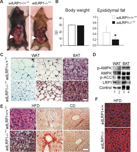
FIGURE 5.
Adipocyte-selective LRP1 knock-out mice exhibit decreased epididymal fat and hepatosteatosis. A, aspect (arrows) and (B), quantification of the epididymal fat and body weight in 20-week-old tamoxifen-treated aP2-CreERT2; LRP1flox/flox;LDLr(-/-) mice (adLRP1(-/-)Tam). C, H&E staining of epididymal WAT and BAT sections from 12-week-old tamoxifen-treated aP2-CreERT2;LRP1flox/flox;LDLr(-/-) mice (adLRP1(-/-)Tam) and tamoxifen-treated controls (aP2CreERT2;LRP1flox/flox;LDLr(-/-)) mice (adLRP1+/+Tam) fed 5 weeks with a high fat diet. D, immunoblot analysis of p-AMPK, AMPK, p-ACC, and loading control in epididymal WAT and BAT, and of LRP1 in BAT and purified epididymal WAT adipocytes from adLRP1+/+Tam (lanes 1, 3) and adLRP1(-/-)Tam (lanes 2, 4) mice. E, H&E (left) and Oil Red O (middle and right) staining of liver sections from adLRP1(+/+)Tam and adLRP1(-/-)Tam mice fed 5 weeks with a high fat diet (HFD) or a regular chow diet (CD). F, Oil Red O staining of liver sections from aP2Cre; LRP1flox/flox;LDLr(+/+) mice. Panels show adLRP1+/+ (aP2Cre-; LRP1flox/flox; LDLr(+/+)) and adLRP1(-/-) (aP2Cre+; LRP1flox/flox;LDLr(+/+)) mice that had been fed a high fat diet (HFD) for 4 weeks. Results are means ± S.D. *, p < 0.05. Scale bar, 50 μm.

