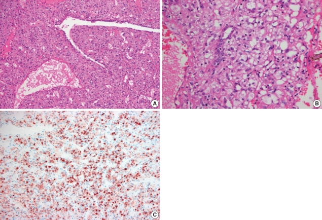
Fig. 3.
Representative hematoxylin and eosin stained sections of the lung mass at low magnification (A) and at high magnification (B). The tumor cells arranged in sheets or trabuculae separated by various sized thin-walled blood vessels. In addition, the tumor cells were polygonal with abundant clear to eosinophilic cytoplasm and distinct cytoplasmic membranes. In the immunohistochemical studies, the tumor cells showed strong immunoreactivity for HMB-45 (C).

