
Figure 3. Expression of Kir channel subunit proteins in native human RPE and neural retina.
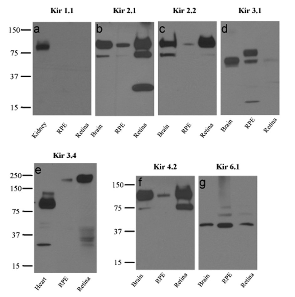
Western blots of human RPE, neural retina, and control tissue lysates probed with anti-Kir1.1 (a), anti-Kir2.1 (b), anti-Kir2.2 (c), anti-Kir3.1 (d), anti-Kir3.4 (e), anti-Kir4.2 (f), or anti-Kir6.1 (g) antibodies. In control experiments, the specificities of anti-Kir1.1, anti-Kir2.1, anti-Kir3.1, anti-Kir3.4, and anti-Kir4.2 antibodies were confirmed in peptide blocking experiments using proteins from the positive control tissues (not shown).