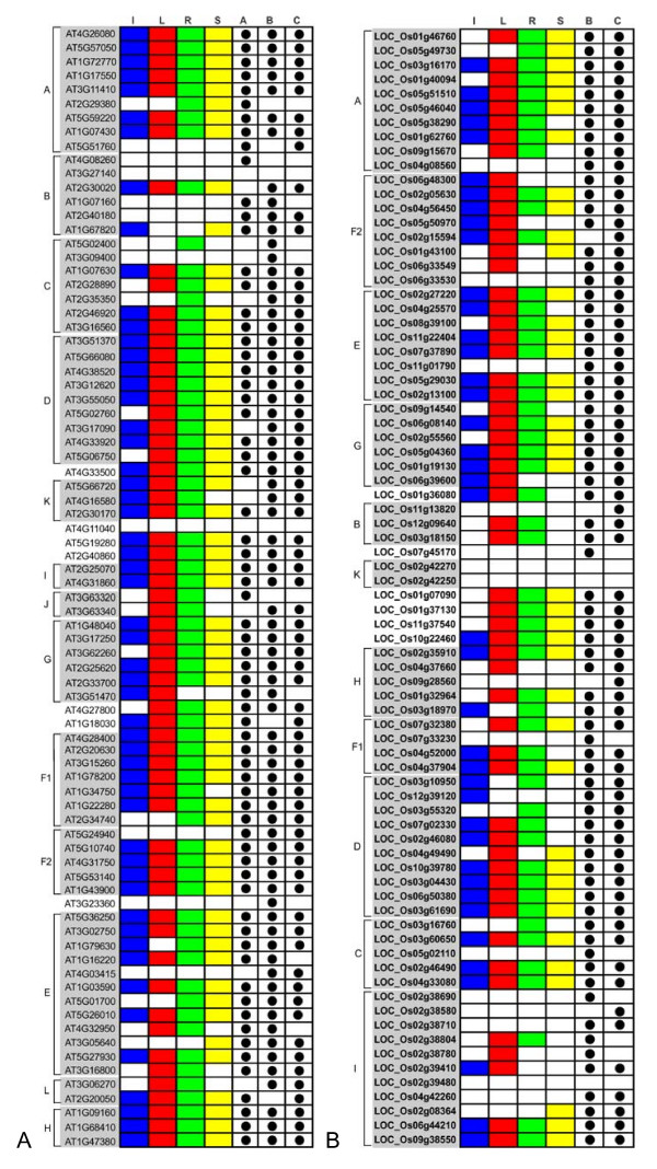
Figure 5.
The expression patterns in different tissues for AtPP2C and OsPP2C genes. Capital letters on the left indicates the major subfamilies. PP2Cs are aligned in the same order as in the corresponding phylogenetic trees. Subfamilies of PP2C genes are highlighted in grey. The expression data of AtPP2C genes in different tissues were combined from microarray (derived from Genevestigator), MPSS, and EST abundance data. The expression data of OsPP2C genes were extracted from MPSS and EST abundance data only. Letters on the top indicate different tissues and databases. A, B and C represent microarray, MPSS and EST data, respectively. A positive signal is indicated by a colored box for the following tissues: blue for inflorescences (I), red for rosette leaves (L), green for roots (R), and yellow for siliques (S). The white boxes indicate that no expression could be detected.

