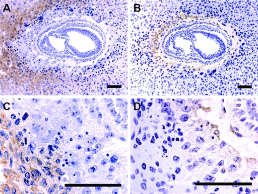
FIG. 1.
CSF-1R is expressed in decidual cells. (A and C) Representative transverse sections of the GD8 uterus of a Csf1op/op mouse immunostained using an antibody against CSF-1R. (B and D) An adjacent section immunostained using an antibody against the trophoblastic cell marker, cytokeratin. Positive cells are stained brown with the antibodies. CSF-1R-positive decidual cells do not colocalize with the cytokeratin-positive trophoblast. Bars = 100 μm.

