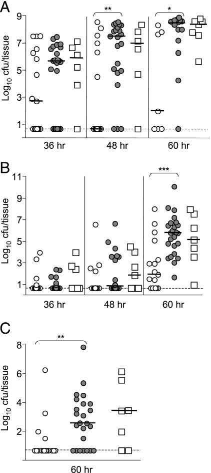
FIG. 2.
Dissemination of the ΔyapE mutant during bubonic infection. Mice were infected subcutaneously in the neck with ∼102 CFU of the ΔyapE mutant (open circles), WT (gray circles), or the yapE complemented mutant (open squares). Mice were sacrificed at 36, 48, and 60 h postinfection, and the colonization of the superficial cervical lymph nodes (A), spleen (B), and lungs (C) was determined. Each symbol represents an individual animal. Black bars correspond to the median CFU/tissue for each group. The dashed line indicates the limit of detection. Asterisks denote significant differences between the median CFU/tissue (Mann-Whitney t test with a two-tailed nonparametric analysis: *, P ≤ 0.05; **, P ≤ 0.005; ***, P ≤ 0.0005). The data represent the composite of three independent experiments. The number (n) of tissues harvested for each indicated time point is given in parentheses in the format “time point = (n)ΔyapE, (n)WT, (n)complemented mutant” as follows: lymph nodes (36 h = 17, 24, 6; 48 h = 17, 22, 6; 60 h = 7, 14, 7), spleen (36 h = 17, 24, 7; 48 h = 17, 24, 7; 60 h = 17, 24, 7), and lungs (60 h = 17, 24, 7).

