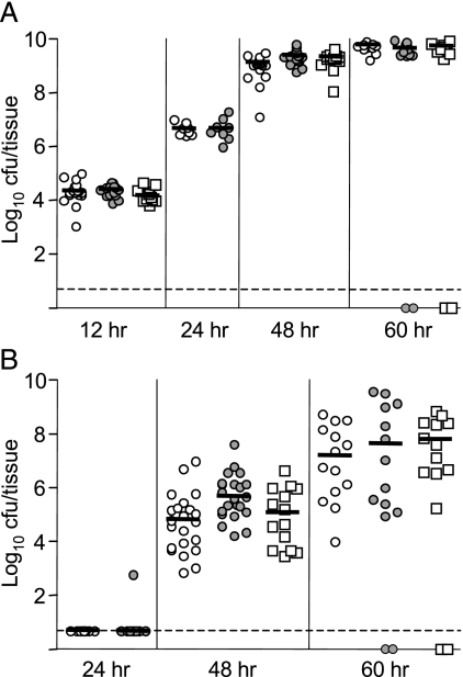
FIG. 3.
Dissemination of the ΔyapE mutant during pneumonic infection. Mice were infected intranasally with ∼105 CFU of ΔyapE mutant (open circles), WT (gray circles), or the yapE complemented mutant (open squares). Mice were sacrificed at 12, 24, 48, and 60 h postinfection, and colonization of the lungs (A) and spleen (B) was determined. Each symbol represents an individual animal, and black bars correspond to the median CFU/tissue for each group. The dashed line indicates the limit of detection. Symbols on the dashed line represent animals with CFU below the limit of detection, and symbols on the x-axis represent animals that succumbed to infection. The number (n) of tissues harvested for each time point is given in parentheses in the format “time point = (n)ΔyapE, (n)WT, (n)complemented mutant” as follows: lungs (12 h = 18, 17, 11; 24 h = 8, 8, 0; 48 h = 21, 20, 14; 60 h = 13, 13, 13) and spleen (24 h = 8, 8, 0; 48 h = 21, 21, 14; 60 h = 12, 14, 12).

