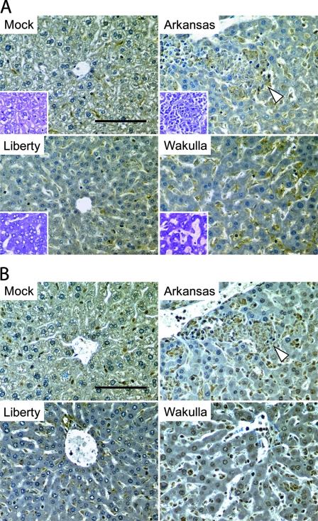
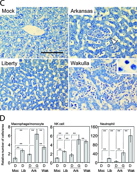
FIG. 1.
Immunohistochemistry of livers infected with three strains of E. chaffeensis. (A) Kupffer cells and macrophages/monocytes in liver specimens from mock- or E. chaffeensis-infected mice immunolabeled with anti-F4/80 antibody. Inserts are hematoxylin-stained livers. (B) NK cells in liver specimens from mock- or E. chaffeensis-infected mice immunolabeled with anti-asialo GM1 antibody. (C) Leukocyte infiltration in liver specimens from mock- or E. chaffeensis-infected mice using Giemsa staining. Insert shows neutrophils at higher magnification (×1,000). (A to C) White arrowheads indicate granulomas. Bar = 100 μm. (D) Relative numbers of macrophages/monocytes, NK cells, and neutrophils per unit area in liver tissues from mock- or E. chaffeensis-infected mice. Cell numbers were determined by counting immuno- or Giemsa-stained cells in five randomly chosen fields (0.79 mm2) each from Liberty (Lib), Arkansas (Ark), and Wakulla (Wak)-infected livers without granuloma (D) or within granulomas (G), and the mean values were normalized to the mean cell numbers from mock (Moc)-infected mice. **, P < 0.01 (ANOVA).


