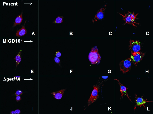
FIG. 4.
Detection of spores and vegetative B. anthracis in RAW 264.7 macrophages by using confocal microscopy. In order to detect spores within infected macrophages, spores of the parent strain and the mutants were labeled with Alexa Fluor 488 (green). Vegetative B. anthracis were visualized by staining DNA with DAPI (blue), which labels DNA from vegetative bacilli and the chromosomal DNA in the macrophage nucleus. Macrophages are highlighted by staining F-actin with Alexa Fluor 568-phalloidin (red). Confocal images display macrophage infections with the B. anthracis parent strain (A to D), the B. anthracis MIGD101 mutant (panels E to H), and B. anthracis ΔgerHA mutant (I to L). Infections are shown with an MOI of 1 after 1 h (A, E, and I), 6 h (B, F, and J), and 12 h (C, G, and K). Infections with an MOI of 10 after 1.5 h are shown in panels D, H, and L.

