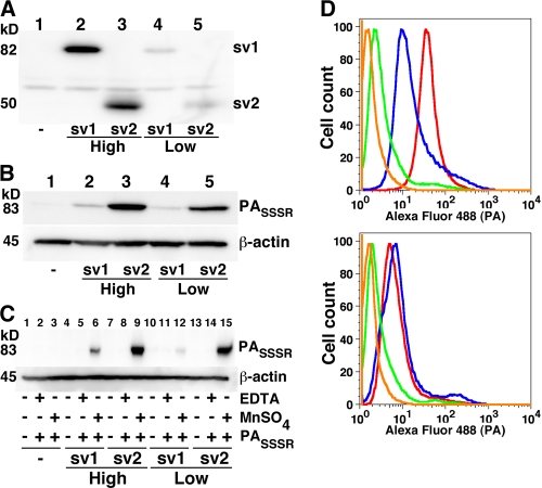
FIG. 1.
Cells that express ANTXR1-sv1 bind less PA than do cells that express ANTXR1-sv2. (A) Receptor-negative CHOR1.1 cells and CHOR1.1 cells stably transfected with either ANTXR1-sv1-HA or ANTXR1-sv2-HA were treated with a non-membrane-permeative biotinylation reagent. Biotinylated proteins were precipitated with streptavidin-agarose and analyzed by Western blotting using anti-HA antibody. (B) Cells were incubated with a furin-insensitive mutant of PA, PASSSR, for 2 h on ice. The cells were washed with PBS and lysed, and the lysates were subjected to Western blotting with anti-PA antibody. Blots were also probed for β-actin to ensure equal loading. (C) Cells were treated with EDTA until they detached from the wells. The cells were then washed with TBS and resuspended in buffer containing either EDTA or MnSO4, as indicated. The cells were incubated with PASSSR for 2 h at 4°C and then washed, and the amount of bound PASSSR was assessed by Western blotting. Blots were also probed for β-actin to ensure equal loading. (D) CHOR1.1 cells (green) and cells stably transfected with ANTXR1-sv1-HA (top panel; blue), ANTXR1-sv2-HA (top panel; red), ANTXR1-sv1-T118A-HA (bottom panel; blue), and ANTXR1-sv2-T118A-HA (bottom panel; red) were incubated with Alexa fluor-labeled PASSSR in suspension for 2 h at 4°C and then washed, and the amount of bound PASSSR was assessed by flow cytometry. As a negative control, cells expressing ANTXR1-sv2-HA were not incubated with Alexa fluor 488-labeled PASSSR (orange).

