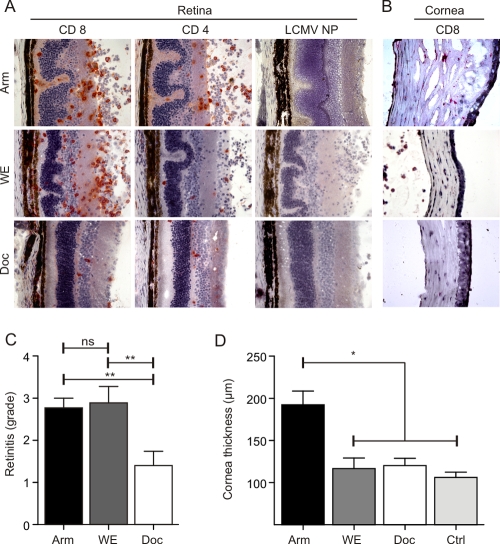
FIG. 1.
In situ analysis of LCMV-induced chorioretinitis and keratitis. C57BL/6 mice were infected i.o. with 103 PFU of LCMV strain Arm, WE, or Docile (Doc). (A and B) Eyes were analyzed by immunohistology on day 12 postinfection for the presence of CD8+ and CD4+ T cells and LCMV nucleoprotein (NP). Representative images of retinas (A) and corneas (B) from three independent experiments are shown. (C) Semiquantitative analysis of immunopathological retinitis following infection with the indicated LCMV strains. Data are mean retinitis scores ± standard errors of the means (SEM; Arm, n = 16 mice; WE, n = 5 mice; Docile, n = 6 mice). (D) Measurement of corneal thickness using digital morphometry. Values are mean corneal thicknesses ± SEM (Arm, n = 13 mice; WE, n = 5 mice; Docile, n = 6 mice; control [Ctrl], n = 10 naïve C57BL/6 mice). **, P < 0.001; *, P < 0.05; ns, not significant.

