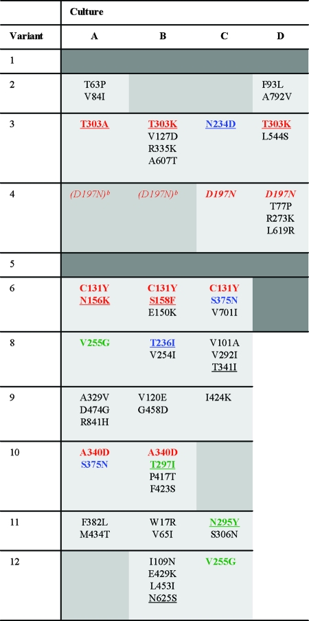
TABLE 1.
Observed amino acid changes in evolved ΔV1/V2 virus variantsa
Amino acid changes that occur in independent cultures containing the same original virus are indicated in red. Substitutions that occur in multiple cultures containing different original viruses are represented in blue. Identical changes that occur in different original viruses in the 8 to 12 cluster are indicated in green. We used the same color code for different variants that result in the loss of the same glycosylation site (N156K and S158F, T303A/K, N295Y and T297I, and N234D and T236I). Mutations that result in the elimination of glycosylation sites are underlined, and mutations that result in the acquisition of a glycosylation site are in italics. Mixed sequences and silent mutations were excluded. The dark gray shading indicates cultures in which we did not find adapted viruses. The medium gray shading indicates cultures in which replicating virus was observed at some point during the 4½ months of culturing but which were lost during cell-free passage.
The D197N mutation was observed in early sequences (data not shown), but the virus was subsequently lost during cell-free passage.

