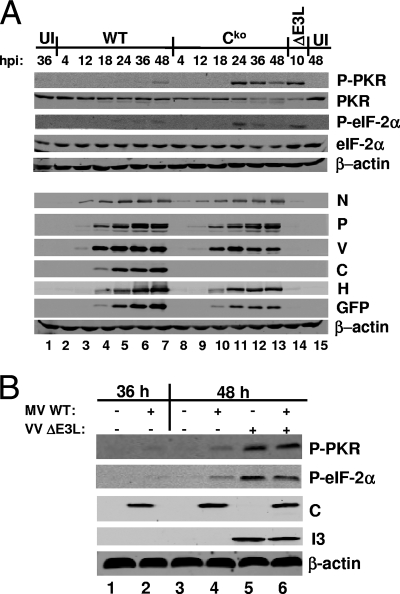
FIG. 3.
PKR and eIF-2α phosphorylation in WT and Cko virus-infected PKR+ cells. (A) Cells were left uninfected (UI) or were infected with WT or Cko MV at an MOI of 5 TCID50/cell or VVΔE3L at an MOI of 5 PFU/cell. Whole-cell extracts were prepared at the indicated times postinfection. Western blot analyses were performed using antibodies against phospho-PKR (P-PKR), PKR, phospho-eIF-2α (P-eIF-2α), eIF-2α, N, P, V, C, H, GFP, and β-actin. hpi, hours postinfection. (B) Cells were left uninfected or were infected with WT MV at an MOI of 5 TCID50/cell. At 38 h after infection with MV, the cells were superinfected with VVΔE3L at an MOI of 5 PFU/cell where indicated (+). Whole-cell extracts were prepared at the indicated times and analyzed by Western blotting with antibodies against phospho-PKR, phospho-eIF-2α, β-actin, MV C, and VV I3. +, present; −, absent.

