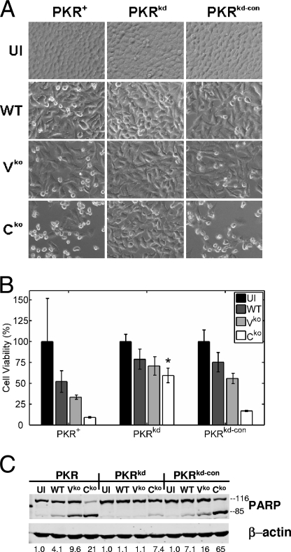
FIG. 4.
MV-induced apoptosis is impaired in PKR-deficient cells. PKR+, PKRkd, and PKRkd-con cells were infected with WT, Vko, or Cko virus or left uninfected (UI). (A) Phase-contrast images taken at 36 h postinfection. (B) Results of the colorimetric MTT assay to measure cell viability 48 h postinfection, displayed as percentages of the number of viable uninfected cells (n = 4). * , P of <0.05 by Student's t test for comparison of the Cko virus yields in PKRkd cells and those in PKR+ or PKRkd-con cells. (C) Western immunoblot analyses performed on whole-cell extracts prepared at 36 h postinfection, using antibodies against human PARP and β-actin. The quantity of PARP cleavage based on the immunoblots, expressed as a ratio of PARP85 to total PARP [PARP85 ÷ (PARP85 + PARP116)], is shown below each lane.

