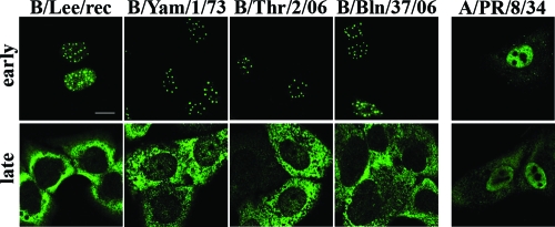
FIG. 1.
The B/NS1 proteins accumulate in a dot-shaped pattern in the nucleus early in infection. Human lung epithelial A549 cells were infected with the influenza B virus strains B/Lee, B/Yamagata/1/73 (B/Yam/1/73), B/Thuringen/2/06 (B/Thr/2/06), and B/Berlin/37/06 (B/Bln/37/06) and influenza A virus A/PR/8/34 at an MOI of 2. Infected cells were processed for microscopic analysis at early (4 to 6 h p.i.) or late (16 h p.i.) times of infection. The cells were stained with primary rabbit antiserum specific for the A/NS1 or the B/NS1 protein, which was followed by detection with secondary α-rabbit Alexa 488 antibodies. The cells were analyzed by CLSM using the 488-nm laser setting. Scale bar = 10 μm.

