FIG. 2.
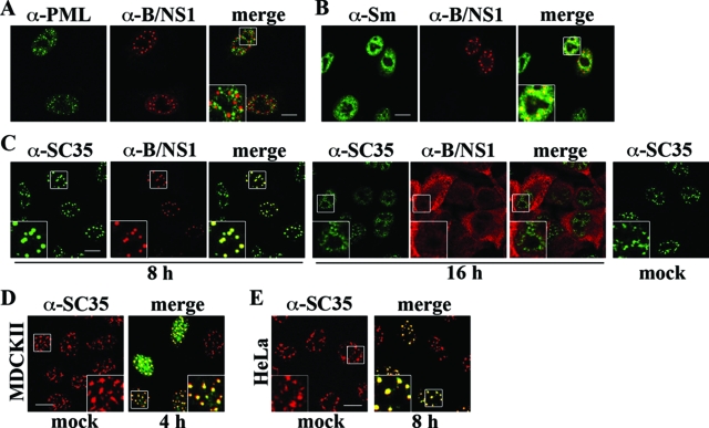
The B/NS1 protein colocalizes with the splicing factor SC35 in nuclear speckles, leading to a coalesced appearance of these domains early in infection. A549 cells were mock infected or infected with influenza B/Yam/1/73 virus for 8 (A, B, and C) or 16 (C) h. Subsequently, the cells were fixed, permeabilized, and double stained with B/NS1-specific rabbit serum (α-B/NS1), together with monoclonal antibodies recognizing either PML bodies (A, α-PML), snRNPs (B, α-Sm), or the speckle marker antigen SC35 (C, α-SC35), followed by detection with secondary α-rabbit IgG-Alexa 594 and α-mouse IgG-Alexa 488 antibodies, respectively. MDCKII cells (D) and HeLa cells (E) were mock infected (left) or infected with influenza B/Lee/rec virus (right). Cells were stained for SC35 only (D and E, mock) or the SC35 (red signal) and B/NS1 (green signal) proteins together (D and E, virus) as described for panel B. The stained cells were analyzed by CLSM. Scale bar = 10 μm. The insets show enlargements of the indicated areas (white squares).

