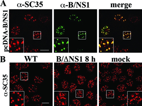
FIG. 3.
The B/NS1 protein accumulates in nuclear speckles in the absence of other influenza B virus proteins. (A) MDCK cells were transfected with the expression plasmid pcDNA3-B/NS1; 36 h posttransfection, the cells were fixed, permeabilized, and stained for the B/NS1 and SC35 proteins. (B) MDCK cells were either mock infected (mock) or infected with WT influenza B virus (WT) or ΔNS1 virus (B/ΔNS1) at an MOI of 5 to infect all cells. The cells were fixed at 8 h p.i. and stained for the SC35 protein, and micrographs were captured by CLSM using the 488-nm and 594-nm laser settings. Scale bar = 10 μm. The insets show enlargements of the indicated areas (white squares).

