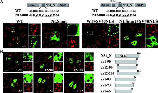
FIG. 8.
Speckle association of the B/NS1 protein requires an intact NLS and amino acids 1 to 90. (A) The schematic drawings at the top show the primary structure of the inserted B/NS1_N polypeptide within the constructed β-Gal-GFP fusion genes. The sequences of the B/NS1 WT and mutant NLS (NLSmut) and the position of the additional SV40 T-Ag NLS are also given. The coding sequences of B/NS1_N (amino acids 1 to 104) carrying a WT or mutant NLS were inserted into the vectors pHM829 and pHM839, respectively, thereby generating β-Gal-GFP fusion proteins of B/NS1_N without (left) or with (right) an additional SV40 T-Ag NLS. MDCK cells were transfected with plasmids expressing the indicated fusion proteins and were stained with SC35-specific antibody and secondary α-mouse IgG-Alexa 594 conjugate. The GFP and Alexa 594 signals were analyzed by CLSM, and fields with merged signals are shown. The white squares in the larger micrographs indicate areas that are expanded on the right, with separate recordings of both channels shown. (B) The scheme on the right presents the structures and lengths of the constructed B/NS1_N derivatives that were fused to the coding sequences for β-Gal and GFP. MDCK cells were transfected with the indicated expression plasmids; 24 h posttransfection, the cells were fixed, permeabilized, and stained with SC35-specific antibody and secondary α-mouse IgG-Alexa 594 conjugate. The GFP and Alexa 594 signals were analyzed by CLSM, and fields with merged signals are shown. The white squares in the large micrographs indicate areas that are expanded on the right, with separate recordings of both channels shown. Scale bars = 10 μm.

